Dosimetry for interventional oncology
Confirm tumour coverage before every ablation.
hapchot generates a patient-specific simulation of your ablation zone.
Confirm coverage. Check margins. Then trigger.
The challenge
Percutaneous tumour ablation is performed blind.
No real-time visualisation of the treated zone. No patient-specific modelling. Practitioners rely on theoretical ablation zones that do not account for individual anatomy or tissue properties.
The curative potential of ablation is not limited by the technique, but by the absence of control over the delivered dose.
* For irreversible electroporation (IRE) of hepatocellular carcinoma (HCC) tumours
Introducing hapchot
The product
Peri-operative assistance for percutaneous tumour ablations
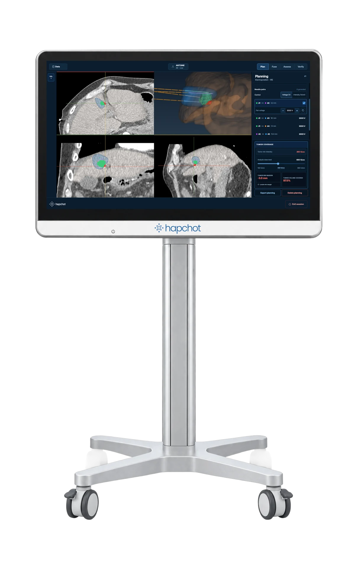
Patient-specific simulation of the ablation zone to confirm tumour coverage and correct needle placement before triggering treatment.
Thermal
Heat or cold destroys the tumour
Electroporation
An electric field destroys the tumour
Radiologists
Intervene with certainty: confirm the margins and adjust the needles before the trigger.
Hospitals
Fewer repeat ablations: better tumour coverage means fewer retreatments.
Patients
Precise treatment from the first procedure: less therapeutic escalation, fewer repeated procedures.
How it works
Plan. Place. Treat. Confirm.
hapchot adds the check before you trigger.
Plan
1 / 6
Plan the ablation
Today
Planned on pre-operative imaging, often with the manufacturer’s theoretical ablation zones.
With hapchot
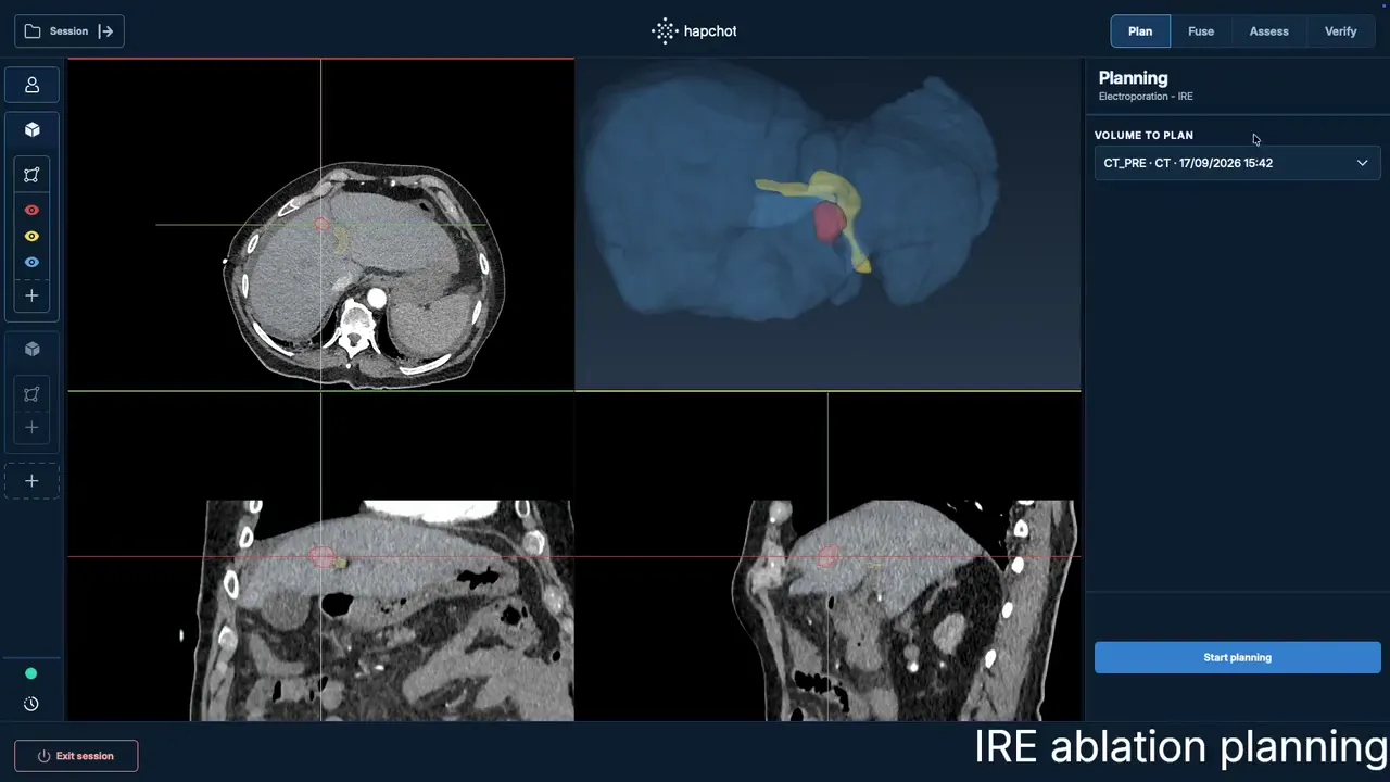
Plan on the patient’s segmented CT or MRI: choose the modality, position the needles or applicators, simulate the zone against the tumour and adjust until the planned coverage and margins satisfy you.
- Electroporation
- Patient-specific ablation zone.
- Thermal
- Theoretical ablation zone. Patient-specific modelling planned for v2.
Place
2 / 6
Place the needles
Today
Needles or applicators are placed under CT or ultrasound guidance, navigation or robotics.
With hapchot
Unchanged. hapchot works with your guidance system.
Check 1/2 · Added by hapchot
3 / 6
Rebuild the anatomy as it is now
Today
Before the trigger, coverage is usually judged by eye on the needle-in images.
With hapchot
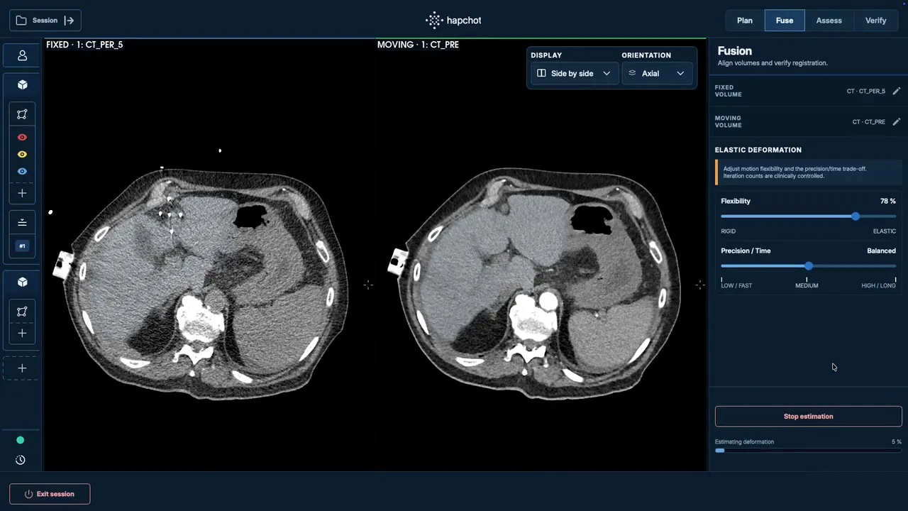
With the needles in place, elastic image fusion rebuilds the anatomy as the patient lies on the table: a digital twin that shows where the tumour and its surrounding anatomy actually are now.
Check 2/2 · Added by hapchot
4 / 6
Check coverage, adjust, re-run
With hapchot
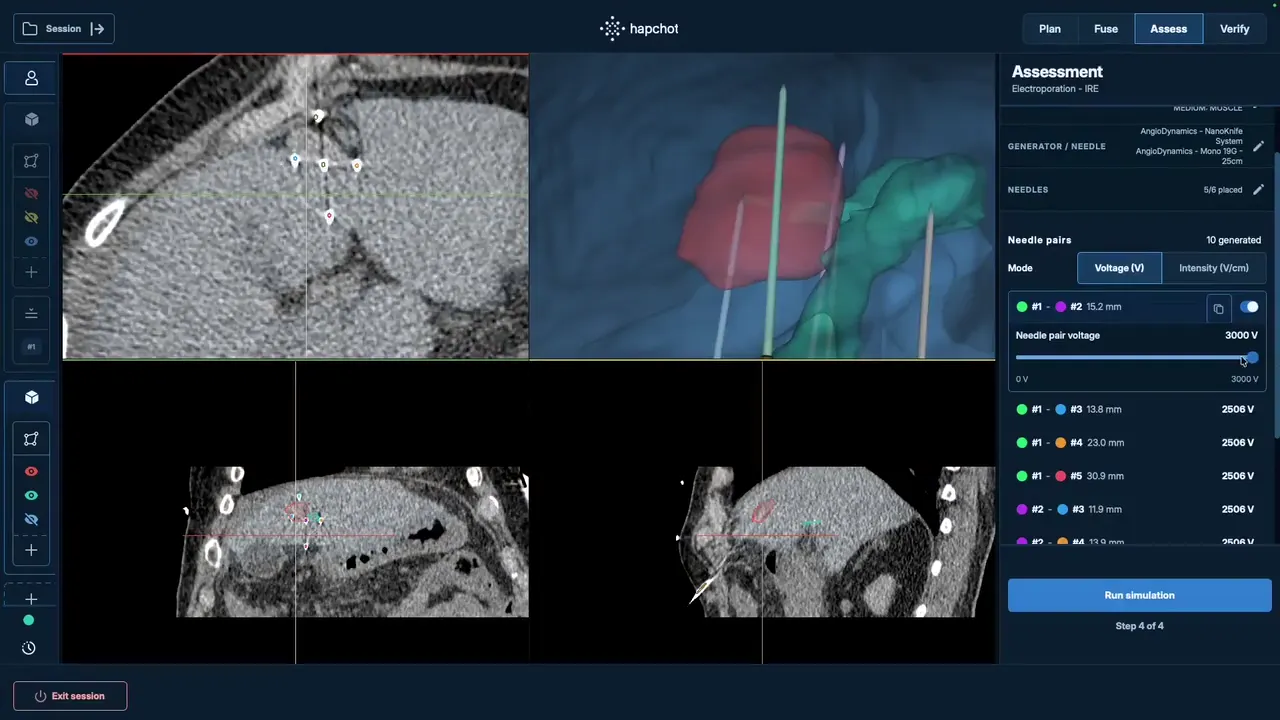
The ablation zone is simulated for the needles as placed and checked against the tumour. If coverage or margins fall short, correct a position and re-run, before any energy is delivered.
- Electroporation
- Patient-specific prediction of the treated zone.
- Thermal
- Theoretical zone on the fused, needle-in anatomy. Patient-specific modelling planned for v2.
Treat
5 / 6
Deliver the energy
Today
Your usual generator delivers heat, cold or an electric field.
With hapchot
Unchanged. hapchot works alongside your generator.
Confirm
6 / 6
Confirm the ablation
Today
Post-ablation imaging, then follow-up imaging.
With hapchot
Register the post-ablation scan with the simulated zone and compare it with the tissue actually treated, to report the margin achieved.
- Electroporation
- Compared with the oedema on a day-3 control MRI.
- Thermal
- Compared with the necrosis on the post-ablation scan, often on the table.
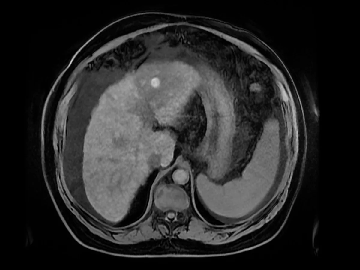
Post-op MRI 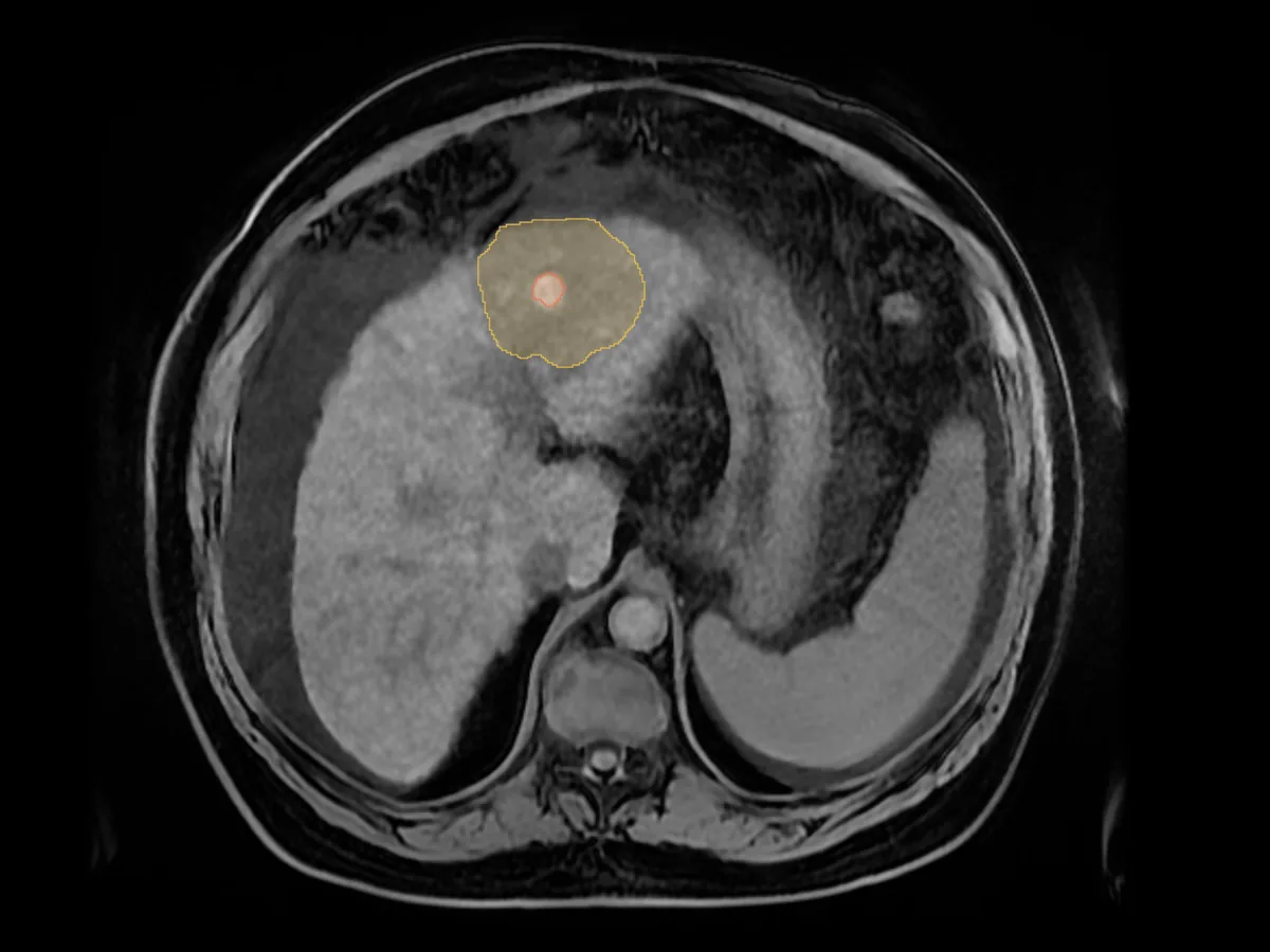
Oedema and tumour segmented
Your workflow stays the same. Now you know the coverage before you trigger, not after.
Hardware-agnostic. Will be available for multiple solid tumour indications.
Developed in collaboration with INRIA, CNRS, AP-HP, Gustave Roussy, CHU Poitiers, and Université Sorbonne Paris Nord






Roadmap
We’re building the product.
The modelling has been validated on 31 patients and is in clinical routine at Hôpital Avicenne.
Building hapchot v1
- Prototype tested by partner centres
- CE technical file in preparation
Conformity and CE submission
- CE submission - hapchot v1
- hapchot v2 R&D - thermal modelling and guidance
CE marking, European launch
- CE-marked and commercially available
- Prospective clinical trial of hapchot v1
- hapchot v2 product development
v2, North America, urology
- hapchot v2 CE-marked
- FDA 510(k): US launch
- hapchotURO: prostate ablation
hapchot v1 — elastic image fusion, patient-specific electroporation modelling, and theoretical thermal ablation zones.
hapchot v2 — adds patient-specific thermal modelling and needle guidance.
Founding team
Product development, science, clinical, and business expertise
Leadership

Luc Lafitte
CTO
Leads
Tech & compliance
Brings
12 years in software development
Lead developer of the hapchot software

Simon Revelly
CEO
Leads
Business, strategy & product
Brings
8 years in medtech, across product and hospital sales
Ex-head of Product @ Pollen Robotics

Scientific and medical advisors

Dr. Baudouin de Senneville
Scientific advisor
Research director @ CNRS
Expert in AI and image processing

Pr. Olivier Seror, MD
Medical advisor
Head of Interventional Radiology @ Hôpital Avicenne, APHP
Global opinion leader in interventional radiology

Dr. Olivier Sutter, MD
Medical advisor
Interventional Radiologist @ Hôpital Avicenne, APHP
Expert in percutaneous tumour ablation
Pr. Olivier Seror, MD
Medical advisor
Head of Interventional Radiology @ Hôpital Avicenne, APHP
Global opinion leader in interventional radiology
Dr. Olivier Sutter, MD
Medical advisor
Interventional Radiologist @ Hôpital Avicenne, APHP
Expert in percutaneous tumour ablation
Clinical partners
Partner with us
We're selecting interventional radiology centres to join research projects around hapchot. hapchot is an investigational device: it is not CE marked and not available for sale.
References
- [1]Sutter O., et al. "Towards Perioperative, Numerically-Assisted Irreversible Electroporation for Hepatocellular Carcinoma: Clinical Outcomes Informed by Numerical Simulations" (2025).DOI: 10.1007/s00330-025-12223-7
Presented at World Congress of Electroporation 2024, Rome — and Journées Françaises de Radiologie 2024, Paris.
- [2]Stättner S., et al. "Ablative margins in percutaneous thermal ablation of hepatic tumors: a systematic review." Expert Review of Anticancer Therapy, 2023.
- [3]Charalel R., et al. "Short-Term Out-of-Pocket and Total Costs of Care After Ablation, Resection, or Transplant for Early-Stage Hepatocellular Carcinoma: A National SEER-Medicare Cost Comparison." AJR, vol. 223, 2024.
- [4]Paolucci I., et al. "International multisociety Delphi consensus for liver tumour thermal ablation: margin assessment." Lancet Oncology, 2026.
Endorsed by ESSE, CIRSE, and SIO. 72-expert Delphi consensus across North America, South America, Europe, and Asia.
- [5]Laimer G., et al. "International multisociety Delphi consensus for liver tumour thermal ablation: procedural and practice standards." Lancet Oncology, 2026.